 پرشین فایل | مرجع دانلود فایل
توضیحات

این فایل در قالب پاورپوینت و در 40 اسلاید قابل ویرایش و به زبان انگلیسی تهیه و تقدیم می گردد.
این فایل جهت همکاران پرستار شاغل و اساتید گرامی و دانشجویان محترم کارشناسی ارشد و کارشناسی پرستاری جهت تدریس و برگزاری کارگاه آموزشی با مبحث الکتروکاردیوگرام پایه به همراه تصاویر متعدد آموزشی و مباحث پایه ای ECG تهیه گردیده است .
بخشی از مطالب این فایل در زیر تقدیم می گردد .
Electrocardiography
•A recording of the electrical activity of the heart over time
• Gold standard for diagnosis of cardiac arrhythmias
• Helps detect electrolyte disturbances (hyper- & hypokalemia)
• Allows for detection of conduction abnormalities
• Screening tool for ischemic heart disease during stress tests
• Helpful with non-cardiac diseases (e.g. pulmonary embolism or hypothermia
Types of ECG Recordings
•Bipolar leads record voltage between electrodes placed on wrists & legs (right leg is ground)
•Lead I records between right arm & left arm
•Lead II: right arm & left leg
•Lead III: left arm & left leg
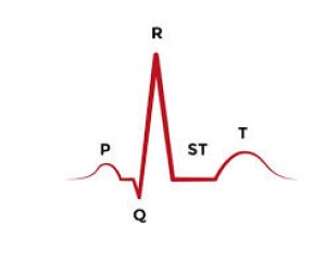
ST segment:
• Connects the QRS complex and T wave
• Duration of 0.08-0.12 sec (80-120 msec
T wave:
• Represents repolarization or recovery of ventricles
• Interval from beginning of QRS to apex of T is referred to as the absolute refractory period
QT Interval
• Measured from beginning of QRS to the end of the T wave
• Normal QT is usually about 0.40 sec
• QT interval varies based on heart rate
Ischemic Heart Disease
•Detectable by changes in S-T segment of ECG
•Myocardial infarction (MI) is a heart attack
•Diagnosed by high levels of creatine phosphate (CPK) & lactate dehydrogenase (LDH)
راهنمای خرید:
- لینک دانلود فایل بلافاصله بعد از پرداخت وجه به نمایش در خواهد آمد.
- همچنین لینک دانلود به ایمیل شما ارسال خواهد شد به همین دلیل ایمیل خود را به دقت وارد نمایید.
- ممکن است ایمیل ارسالی به پوشه اسپم یا Bulk ایمیل شما ارسال شده باشد.
- در صورتی که به هر دلیلی موفق به دانلود فایل مورد نظر نشدید با ما تماس بگیرید.
دیدگاهها
هیچ دیدگاهی برای این محصول نوشته نشده است.